搜索
| 咨询热线:18511236489 |
Layer V Glutamatergic Neurons
Layer V Glutamatergic Neurons

rigin: Human iPSC line
Products
5M Viable Cells/Vial - Standard (BX-0350)
2M Viable Cells/Vial - GFP (BX-0351)
Contents: 1 vial of cells and corresponding BrainXell supplements
Specifications
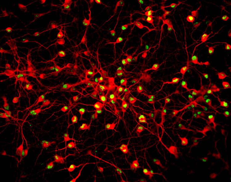
Marker Expression: Layer V Glutamatergic Neurons (BX-0350) have high neuronal purity (>90%) and comprise predominantly excitatory neurons. Labeling with the pan-neuronal marker MAP2 (red) and the layer V specific marker CTIP2 (green) highlight the purity of layer V glutamatergic neurons in this product.
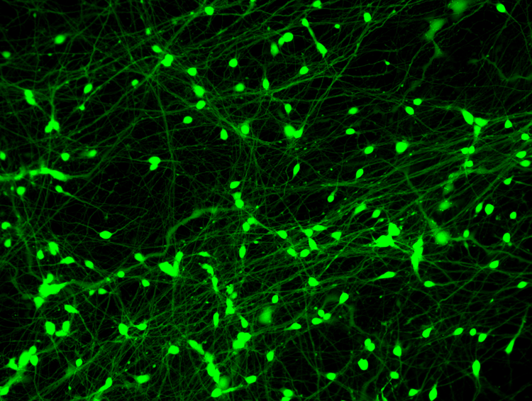
Morphology: Layer V Glutamatergic Neurons (BX-0350) exhibit substantial neurite outgrowth within a week in culture and are adherent. Calcein staining (green) demonstrates the characteristic oval cell shape and long processes of Layer V Glutamatergic Neurons in culture.
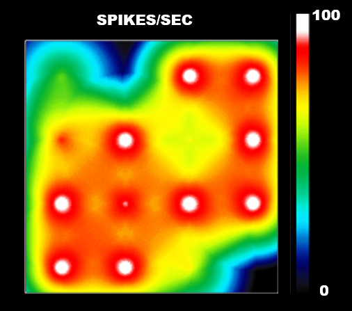
Function: Layer V Glutamatergic Neurons (BX-0350) exhibit pronounced electrophysiological activity after two weeks in culture, as demonstrated by multi-electrode array (MEA) recordings.
Applications
Calcium Influx Assays:
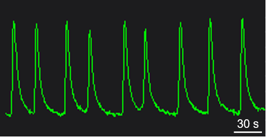
Changes in calcium concentration are closely tied to neuronal activity as action potentials are associated with large pre-synaptic calcium influx and a notable rise in postsynaptic calcium at excitatory synapses. This can be observed experimentally by stimulating the neurons or culturing the neurons under suitable conditions to form mature networks that exhibit spontaneous oscillations. The influx of calcium can be measured using a variety of calcium-sensitive fluorescent dyes, which are commercially available.
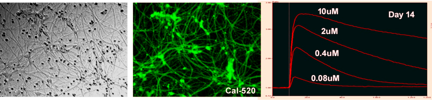 After 14 days in culture Layer V glutamatergic neurons show a strong calcium response evoked by applications of 0.08 µM to 10 µM glutamate.
Layer V Glutamatergic Neurons (BX-0350) were cultured in 96-well plates for three weeks and then loaded with Calbryte-520. Spontaneous oscillations were recorded in all wells simultaneously using an FDSS/µCell Functional Drug Screening System (Hamamatsu).
MEA Assays:
Multi-electrode arrays (MEA) measure extracellular voltage changes that occur as neurons fire action potentials. These measurements reveal the firing patterns of individual neurons as well as the patterns of neuronal networks that exist in the cell culture. Such measurements are non-invasive and allow for repeated recordings.
Layer V Glutamatergic Neurons (BX-0350) were cultured on Axion Biosystems MEA plates for several weeks and recorded regularly. Below, a time course of the number of active electrodes, mean firing frequency, and synchrony index reveal the development of neuronal activity in Layer V Glutamatergic Neurons over several weeks in culture.
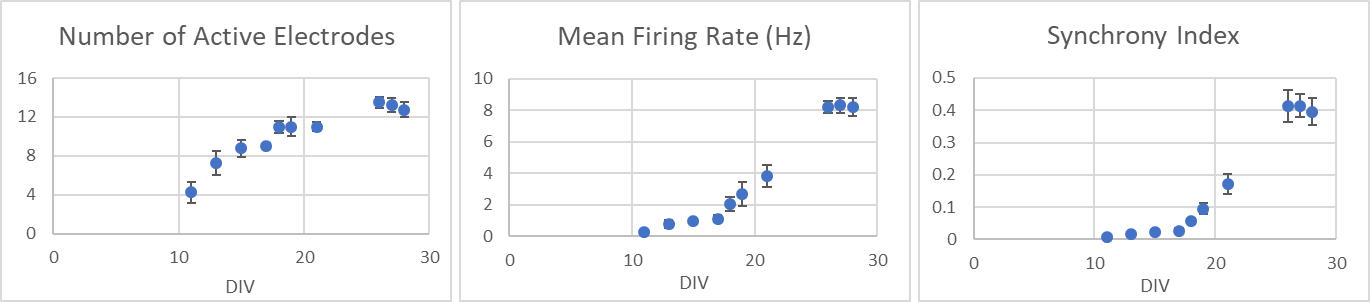
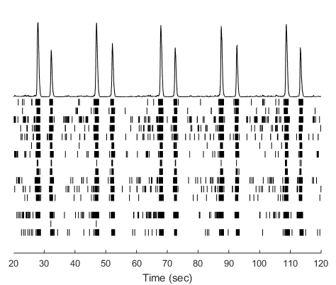
The raster plot of spike activity shows network bursting observed on day 24 for Layer V Glutamatergic Neurons.